Computerized Tomography (CT) vs. Magnetic Resonance Imaging (MRI) Scans for Diagnosing Strokes in Adults 65 Years and Older: A Comparative Analysis
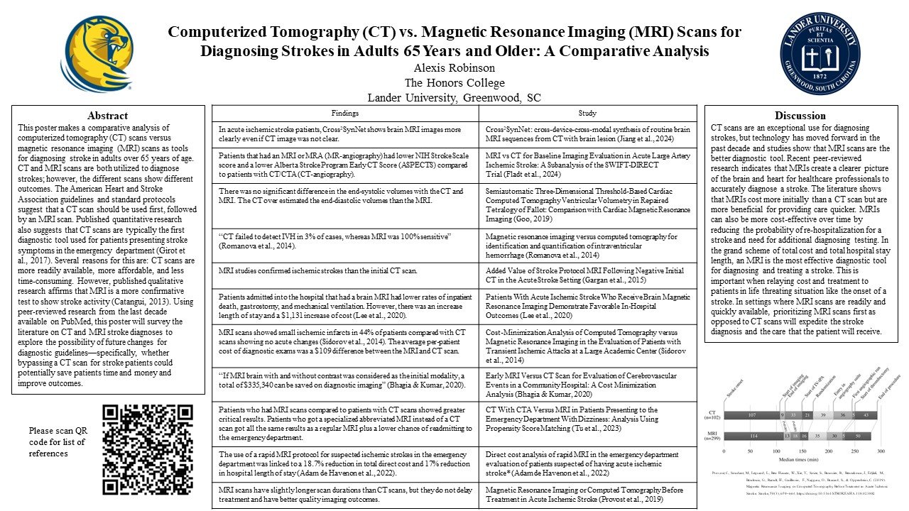
This poster makes a comparative analysis of computerized tomography (CT) scans versus magnetic resonance imaging (MRI) scans as tools for diagnosing stroke in adults over 65 years of age. CT and MRI scans are both utilized to diagnose strokes; however, the different scans show different outcomes. The American Heart and Stroke Association guidelines and standard protocols suggest that a CT scan should be used first, followed by an MRI scan. Published quantitative research also suggests that CT scans are typically the first diagnostic tool used for patients presenting stroke symptoms in the emergency department (Girot et al., 2017). Several reasons for this are: CT scans are more readily available, more affordable, and less time-consuming. However, published qualitative research affirms that MRI is a more confirmative test to show stroke activity (Catangui, 2013). Using peer-reviewed research from the last decade available on PubMed, this poster will survey the literature on CT and MRI stroke diagnoses to explore the possibility of future changes for diagnostic guidelines—specifically, whether bypassing a CT scan for stroke patients could potentially save patients time and money and improve outcomes.
Alexis Robinson is a public health major from Irmo. Alexis served as a President Ambassador and a LINK peer leader, and she was a resident assistant at Centennial Hall, Lide, and McGhee Court. A member of the Honors College and Lander Leadership Program, Alexis interned for Cornerstone, a substance abuse treatment organization, as well as for the Clemson Extension.